
目次
上腕二頭筋長頭・短頭
(Biceps brachii muscle)
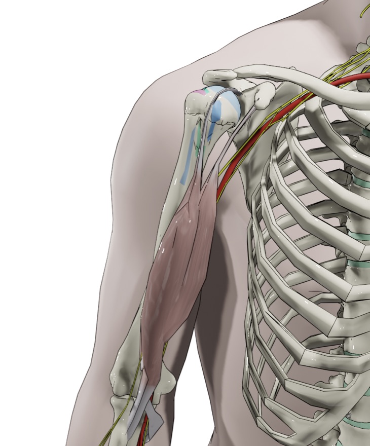
起始(Origin)
短頭(Short head):肩甲骨の烏口突起
(Coracoid process)
長頭(Long head):関節上結節(Supraglenoid tubercle)
、後上方関節唇[12]

停止(Insertion)
橈骨粗面(Radial tuberosity)
Bicipital aponeurosis


上腕二頭筋は起始から90度外旋して停止します[8]。橈骨粗面上の付着部は三日月状で、長さは約21mm、幅は約7mmです[7,8]。長頭は橈骨粗面の近位尺側に付着し、短頭は遠位尺側に付着します。
Footprintの面積は平均108 mm²で、長頭が平均48mm²、短頭が平均60mm²を占めます[8]。

橈骨粗面への停止腱は上腕二頭筋の長頭短頭どちらも寄与しますが、 Bicipital aponeurosisは上腕二頭筋短頭から伸びます。Bicipital aponeurosisが肥厚すると正中神経を絞扼する可能性があります[5]。
支配神経(Innervation)
筋皮神経(C5,C6)
(Musculocutaneous nerve)
栄養血管(Blood supply)
上腕動脈
(Brachial artery)
鎖骨下動脈の延長線上にある腋窩動脈は大円筋/広背筋の下縁から上腕動脈となり肘前部の直下まで伸び、肘窩(cubital fossa)に入った後、橈骨頚部(radial neck)の高さで尺骨動脈と橈骨動脈に分かれます[1,2,3,4,13]。
最初は上腕骨の内側にありますが、徐々に上腕骨の前方に向かって走行していきます。
作用(Action)
前腕の屈曲(Flexion)、回外(Supination)
解剖学的変異
(Anatomical variations)
上腕二頭筋の解剖学的変異はめずらしくなく、Third headが見られることがあります[9,10,11]。Third headの起始部はバリエーションがあり、大胸筋や上腕二頭筋短頭、上腕骨内側に起始したりと様々です。
Third headが正中神経を絞扼する可能性もあります[9]。
←単語帳に戻る
References
[1]Hansen, J., Netter, F. & Machado, C. (2019). Netter's clinical anatomy. Philadelphia, PA: Elsevier.
[2]Sinnatamby, C. & Last, R. (2011). Last's anatomy : regional and applied. Edinburgh New York: Churchill Livingstone/Elsevier.
[3]Fehringer, E., Lippitt, S., Matsen, F., Rockwood, C., Sperling, J. & Wirth, M. (2017). Rockwood and Matsen's The shoulder. Philadelphia, PA: Elsevier.
[4]Standring, S. (2016). Gray's anatomy : the anatomical basis of clinical practice. Philadelphia: Elsevier Limited.
[5]Caetano EB, Vieira LA, Almeida TA, Gonzales LAM, Bona JE de, Simonatto TM. Bicipital aponeurosis. Anatomical study and clinical implications. Rev Bras Ortop. 2018;53(1):75-81.
[6]Szewczyk B, Polguj M, Paulsen F, et al. A proposal for a new classification of coracobrachialis muscle morphology. Surg Radiol Anat. 2021;43(5):679-688.
[7]Forthman CL, Zimmerman RM, Sullivan MJ, Gabel GT. Cross-sectional anatomy of the bicipital tuberosity and biceps brachii tendon insertion: relevance to anatomic tendon repair. J Shoulder Elbow Surg. 2008;17(3):522-526.
[8]Athwal GS, Steinmann SP, Rispoli DM. The distal biceps tendon: footprint and relevant clinical anatomy. J Hand Surg Am. 2007;32(8):1225-1229.
[9]Yershov D, Hudák R. Unusual variation of the biceps brachii with possible median nerve entrapment. Prague Med Rep. 2015;116(2):167-172.
[10]Nayak SR, Prabhu LV, Sivanandan R. Third head of biceps brachii: A rare occurrence in the Indian population. Ann Anat. 2006;188(2):159-161.
[11]Sargon MF, Tuncali D, Celik HH. An unusual origin for the accessory head of biceps brachii muscle. Clin Anat. 1996;9(3):160-162.
[12]Vangsness C, Jorgenson S, Watson T, Johnson D. The origin of the long head of the biceps from the scapula and glenoid labrum. An anatomical study of 100 shoulders. The Journal of Bone and Joint Surgery British volume. 1994;76-B(6):951-954.
[13]Patnaik, Gopichand & Kalsey, G. & Singla, R.K.. (2002). Branching pattern of brachial artery-A morphological study. J Anat Soc India. 51. 176-186.
注)本記事は一介の臨床家が趣味でまとめたものです。厳密さや正確性は不十分なものとなっており、内容には諸説あります。最終的な情報の判断は個人にお任せします。